


Join us
If you believe in the power of what science can do, join us in our endeavour to push the boundaries of science to deliver life-changing medicines.
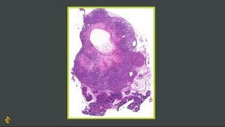
Integral to modern drug discovery is the ability to understand – in as much detail as possible – the effects of our compounds on human tissues at a cellular level. In other words, exactly what our candidate drugs are doing in a patient’s body. For decades, scientists have used the traditional techniques of histology and histopathology, staining tissue samples and looking for particular cellular morphologies, markers and signals on microscope slides. To work out where drug molecules go within the body (biodistribution), previously the only available option was autoradiography, an expensive and laborious radio-labelled approach with limited ability to only image distribution of a single target.
Now, a suite of innovative technologies which make up our advanced molecular imaging capabilities have enabled a quantum leap in our ability to understand disease processes and evaluate drug efficacy and safety. These technologies let us probe every tissue sample – whether from patient biopsies, animal models or advanced cell cultures – in unprecedented depth. Combining these remarkable powers of detection with the incredible analysis and interpretation capabilities of artificial intelligence (AI) and machine learning means we can explore uncharted territory with open minds in our search for the unknown and unexpected.
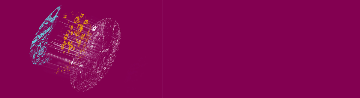
In this field, advances in mass spectrometry imaging (MSI) have been the real game changer. We are now able to simultaneously measure the individual masses of molecules using a mass spectrometer, to visualise their spatial distributions – whether peptides, proteins, lipids, endogenous metabolites or drug molecules – within the microenvironment of a tissue, providing vital clues to their inter-relationships and allowing us to better assess the safety and efficacy of compounds.
As a measurement tool, mass spectrometry has reached unprecedented levels of power, precision and versatility. Its uses span a vast breadth of applications: from oceans to operating theatres to missions on Mars. Now it’s helping us navigate the full molecular complexity of human tissue in health and disease.
Mass spectrometry itself is of course not new. Utilised in many areas of research and development, it relies on the process of ionisation: turning tissue samples into gaseous form. What is special about MSI is that the tissue sample is not homogenised (mixed-up) prior to ionisation. Instead, it is snap-frozen and ionised directly from its intact surface, so that each molecule’s original position is known. The whole sample is scanned a few microns at a time with each discrete location analysed forming one pixel in the images we ultimately generate. This provides a wealth of digital information.
Today’s advanced molecular imaging techniques – and MSI in particular – mean we can now create the most detailed molecular maps ever. Much like Google Earth software enables a satellite view of the planet down to 3D views of individual streets and buildings, our new imaging capabilities allow us to zoom in and out from the micro to the macro level and back. Our mass spectrometers even enable us to do the equivalent of looking through the window of a house to see where the sofa is.
Every molecule we detect has its own map, pieced together by tens of thousands of images from different perspectives. We can “see” drug molecules, biomarkers and the tissue microenvironment simultaneously and examine the picture from the genomic and molecular viewpoint up to the cellular, tissue, organ and patient level. Vast datasets are generated from healthy, diseased and drug-treated tissue samples which can then be mined by AI and machine learning techniques to spot patterns, connections and relationships at a level of complexity far greater than ever before, turning information into insights and insights into knowledge.
Armed with this knowledge we’ll be more equipped to design safe and effective drugs, develop optimal drug delivery methods, work out appropriate dosing and monitor disease progression. Using these approaches, we are already uncovering new insights such as:
AstraZeneca is part of an innovative five-year multi-centre, multidisciplinary collaboration that aims to plot the most detailed map ever seen of the molecular landscape of malignant tumours.
Funded by Cancer Research UK (CRUK), this ‘Grand Challenge’ is led by Professor Josephine Bunch from the National Physics Laboratory and the consortium includes leading scientists from the Francis Crick Institute, the Beatson Institute in Glasgow, the University of Cambridge and many others.
Using a variety of novel MSI instruments, we aim to be ‘cartographers of cancer’, creating the most ambitious molecular map in existence and – like the famous ‘Rosetta Stone’ – using machine learning to decode all the secrets from the data we generate.
The team’s database will be made available to researchers worldwide and its approaches used to create standardised, best-practice guidance for deployment of the latest technologies.
Partnering with academia is key for us to push the boundaries of science and drive disease discovery and understanding to help us create the next generation of therapeutics.
If you believe in the power of what science can do, join us in our endeavour to push the boundaries of science to deliver life-changing medicines.
We know that however innovative our science, however effective our medicines and delivery, to achieve all we want to achieve, we cannot do it alone.